Illustrations
Hymenochirus, Cecere (1998)
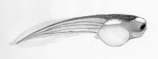
This is my illustration from a living specimen of Hymenochirus at
about 25 days of age. Its overall length is just over 1 cm. A
limb bud can be seen below the tail fin. This is a well-fed tadpole
as indicated by the rounded abdomen. The eyes protrude from the skull
and are angled slightly downward. The mouth is superior and rests between
the eye protrusions. Based on the description in
RABB
(1963) this is a specimen of H. boettgeri. The iridocytes
on this specimen at 25 days were silver but many members of this batch had
golden iridocytes at earlier stages. The longitudinal markings on the
tail are not obvious until week 3.
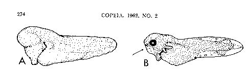
Sokol (1962) Copeia 2:274 "Fig. 1. Young larvae of Hymenochirus. A. H. boettgeri at hatching. Note ventrally produced cement gland. Actual size 2.5 mm. B. H. curtipes 24 hours after hatching. Vertical groove is olfactory pit; horizontal groove marks dorsal rim of developing oral tube (arrow). Actual size 3.5 mm."
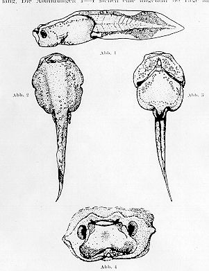
Hymenochirus curtipes, Sokol (1959) Zoologisher Anzeiger 162:156 This illustration is from a paper written in German. I include it here because it is repeatedly referred to in Sokol's1962 paper.
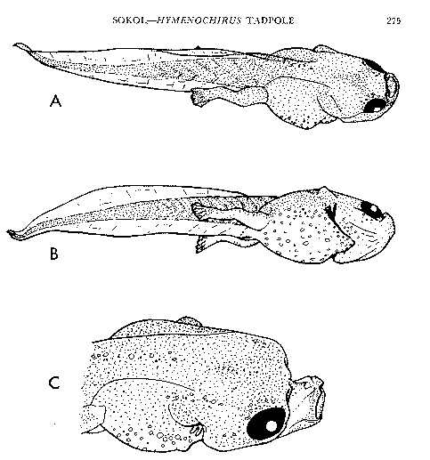
Hymenochirus boettgeri, Sokol (1962) Copeia 2:275 "Fig. 2. Fully grown tadpoles of Hymenochirus boettgeri (21mm). A. Dorsolateral view. B. Ventrolateral view. C. Head of tadpole (18 mm) preserved with mouth open."
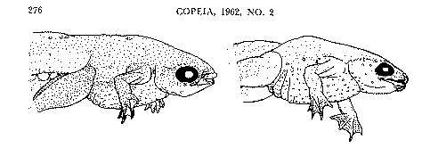
Hymenochirus boettgeri, Sokol (1962) Copeia 2:276
|
||
 Jump to Main